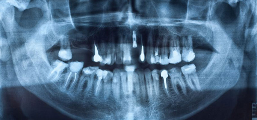
Cone beam computed tomography offer us a window into your teeth and jaws
In the beginning of medical imagery X-rays were considered new and truly innovative to help treat patients. X-rays have been used regularly in the medical profession for decades so every kind of doctor and surgeon can take a look inside the human body without having to make a cut.
The field of radiology has come a long way since the first X-ray image. A variety of new devices and medical imaging methods have been developed and introduced worldwide. One standout to this growing list has turned out to be extremely helpful and beneficial to us and our oral surgery practice: cone beam computed tomography, or CBCT.
Cone beam computed tomography combines the accessibility of old-fashioned X-rays with the raw imaging that modern-day computers can provide. To use the cone beam imaging device, it simply encircles the patient’s teeth and jaws, taking numerous photos using cone-shaped X-rays. The process is completed quickly, and the “volumetric data set” (or large sample of images) is woven together into a single three-dimensional image.
It’s that 3D definition that we need to give us a far better view of a patient’s teeth and jaws. The image can even rotate in any direction as needed to offer more visibility. So preparing to perform dental implant or facial reconstruction procedures is inclusive to this better view prior to surgery. Conditions like oral cysts, a particular pathological process or anything else surgeons need to see are clear as day.
Now, CBCT are in regular use and this includes our office. Improvements and advances in imaging techniques are happening all the time –one recent announcement from a CBCT maker in 2011 declared that its latest device reduced radiation exposure by over 50 percent. (The FDA concluded that radiation from dental CBCT exams are safe, but people under 21 are advised to avoid all forms of radiation exposure unless medically necessary.)
CBCT technology was originally introduced commercially in the early 2000s and it didn’t long to catch on because by 2005, researchers were reporting in medical journals that cone beam computed tomography was here to stay. And we have to agree; cone beam computed tomography may seem like just another chapter in the long history of medical advancements–but for us, it’s one that is greatly enhances our ability to treat patients.

